A versatile organ-on-a-chip approach for in vitro drug testing
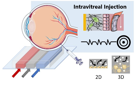
 We propose a versatile approach to develop in vitro models for various parts of the eye, suitable for drug PK and PD testing in a variety of ocular drug delivery routes. Our group is uniquely positioned to develop such technologies: by leveraging our expertise in transport phenomena and microhydrodynamics, we can adapt bulk tissue culturing methods to a microfluidic setting and precisely control the spatiotemporal profile of cell growth and phenotypes; by combining in vitro measurements and quantitative analysis, we can gain detailed knowledge about drug transport across multiple biological barriers compared to existing in vivo and ex vivo methods offering limited outputs. We are currently working with researchers at Mass Eye and Ear to develop this technology, which will fulfill the unmet need for standardized drug testing assays in the fast-growing ocular drug market.
Funding Sources:
 |
 |
 |
 |